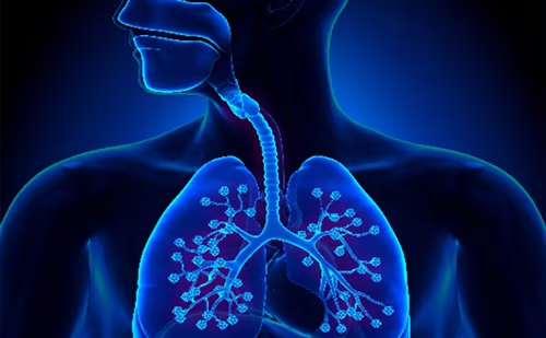
Paediatric sleep-disordered breathing (SDB) refers to a spectrum of respiratory disorders with intermittent upper-airway obstruction and sleep disruption in children.1 SDB spans primary snoring, upper-airway resistance syndrome, obstructive hypoventilation and obstructive sleep apnoea (OSA), listed in order of increasing severity of SDB stratified by polysomnography.2 Paediatric OSA is characterized by recurring episodes of airway obstruction, which fragment sleep and disrupt gas exchange, and is typically measured by the apnoea-hypopnoea index (AHI).3 Although there is no uniformly accepted classification of its severity, OSA is often categorized as mild, moderate or severe based on AHI thresholds of 1, 5 and 10, respectively.4 The primary symptom observed by caregivers in children with SDB is snoring. Other commonly reported symptoms during sleep are gasping, respiratory pauses, restlessness, night sweats and nocturnal enuresis.5
Most children with OSA have enlarged tonsils and adenoids, also called adenotonsillar hypertrophy.6 However, in adults, excessive adipose tissue commonly narrows the upper airway and contributes to upper-airway obstruction.7 While obesity is a risk factor for OSA in children, it is the primary cause of upper-airway narrowing less often than in adults.8 Therefore, the first-line treatment for OSA in children is surgically removing the adenoids and tonsils, which improves or resolves symptoms in most children.4,9 Milder instances of OSA may be managed by intranasal corticosteroids, or simply monitored due to the potential for OSA to spontaneously resolve over time.10
The prevalence of paediatric OSA is 1–4%, though this is likely underestimated as caregivers must identify associated symptoms.11 Untreated OSA is linked with neurobehavioural, cardiovascular and metabolic abnormalities.12 Cognitive and behavioural deficits observed in children with OSA include impaired executive function, inattention, aggression, depression and decreased scholastic performance.5 The intermittent hypoxia (IH) and fragmented sleep associated with recurrent apnoeic episodes may impair cerebral oxygenation, and could account for the neurocognitive deficits in some children.13 Animal models of chronic IH have helped elucidate brain-related structural changes and related neurobehavioural morbidities. These models attempt to replicate the cyclical episodes of hypoxia and reoxygenation observed during apnoeic episodes in children.14 Murine models of IH demonstrate that reactive oxygen species are generated in the cerebral cortex, thereby causing oxidative damage resulting in neuronal cell apoptosis.14 Furthermore, the patterns of inflammation and oxidative stress in IH are similar to those observed in neurodegenerative disorders, demonstrating the potential long-term negative impacts.15
Children often demonstrate a greater burden of neurobehavioural consequences of OSA than adults,16 a finding supported by parallel observations of rats exposed to IH during critical periods of post-natal development. IH was associated with maximal neuronal apoptosis occurring at age 10–25 days, correlating with the peak incidence of upper-airway obstruction in children.17 A follow-up study that exposed juvenile rats to IH throughout this critical period of vulnerability revealed significant deficits on a cognitive spatial-learning test.18 In addition, male juvenile rats showed locomotor hyperactivity, additionally supporting the possibility of sex differences in the manifestations of OSA.18 This could correspond to the greater prevalence of hyperactivity seen in children with OSA,19 although further research is required to elucidate if chronic IH-mediated locomotor hyperactivity is impacted by sex.
The developing brain is vulnerable to injury by the chronic IH seen in OSA. Cognitive and behavioural outcomes of children with OSA are highly variable, and the neural basis of these manifestations is poorly understood.20 Thus, this review explores brain imaging findings of children with OSA described in literature, which may further our understanding of the neurodevelopmental impact of this disorder. Here we describe these imaging findings, largely based on magnetic resonance imaging (MRI)-based studies, organized by macro- and microstructure, function and neuronal metabolism.
Structural magnetic resonance imaging studies of brain macrostructure
Brain morphometry is commonly assessed by MRI to detect gross structural changes.21 Philby et al. revealed grey matter losses in the frontal, parietal and temporal lobes, as well as the brainstem, in 16 children with OSA compared with 200 controls.22 These regions are central in regulating mood and cognition, which explains the clinical features of aggression and cognitive deficits seen in OSA.22 Strengths of the study included the rigorous methodology and the inclusion of 191 controls from the Pediatric Imaging, Neurocognition, and Genetics (PING) data repository.23 The primary weakness was the small sample size of the OSA group. The small sample size was implicated in the lack of correlation between changes in cortical structure and cognitive scores.22
Chan et al. similarly identified lower grey matter volumes in the pre-frontal and temporal regions in eight children with moderate-to-severe OSA when compared with 15 controls.24 These lower volumes also correlated with visual-fine motor coordination deficits, which the authors suggest could be mediated by observed reductions in the lateral occipital gyrus, which is closely related to the visual cortex. These findings were not replicated in 15 children with mild OSA, demonstrating a potential severity-dependent impact of OSA on brain morphometry.24
Macey et al. showed significantly altered cortical thickness in 16 children with OSA compared with 138 controls from the PING study.25 Thinning was observed in the frontal, pre-frontal and parietal cortices, and is consistent with previous findings. Cortical thickening was identified in the bilateral pre-central gyri, the left central gyrus and insular cortices, likely caused by inflammation secondary to hypoxia or disrupted synaptic pruning process.25 There was no correlation between cortical thickness alterations and neurocognitive dysfunction in this study, also attributed potentially to the small sample size.
Musso et al. identified cortical thinning and lower grey and white matter volumes in the frontal, parietal, temporal and occipital areas in 27 children with OSA compared with 18 controls.26 Correlations between neuroimaging findings and cognitive performance were not assessed. Strengths of this study include the large sample size and strict exclusionary criteria to mitigate the impact of covariates such as central obesity. There was no relationship between OSA severity and brain imaging findings.
In the largest population-cohort-based study to date, Isaiah et al.27 examined MRI findings, SDB symptoms and behavioural measures in 10,140 pre-adolescents in the Adolescent Brain Cognitive Development dataset.28 Altered grey matter volume mediated the relationship between SDB symptoms and problem behaviour scores.27 A primary strength of this study was the use of statistical modelling to control confounding from demographic and socioeconomic factors. A weakness was the lack of polysomnography data to corroborate parent-reported symptoms of SDB.
Lv et al. reported lower volumes in the ventral posterior and medial dorsal nuclei of the left thalamus and higher volume in the left pallidum in 25 children with OSA compared with 30 controls.29 The medial dorsal nucleus has reciprocal connections with the pre-frontal cortex and limbic system, and could thus predict deficits in executive function and mood regulation in children with OSA.30
Diffusion magnetic resonance imaging studies of brain microstructure
Diffusion MRI visualizes brain microstructure, quantifying changes in white matter architecture.31 Cha et al. revealed disruptions in hippocampal microstructure in 11 children with moderate-to-severe OSA compared with 12 controls using diffusion tensor imaging (DTI).32 Children with OSA had lower mean diffusivity in the left dentate gyrus, an area of the brain central to verbal learning and memory. The authors, however, did not find macrostructural volumetric changes in grey or white matter, a finding that contradicts the literature. This can potentially be explained by changes in the left dentate gyrus being an earlier consequence of OSA or that the study was underpowered to detect smaller effects of OSA on these brain regions.32
Horne et al. identified both acute and chronic changes when comparing 18 children with SDB with 20 controls.33 Acute changes, characterized by decreased mean diffusivity, were reported in the hippocampus, insula, thalamus and temporal, occipital and cerebellar sites. Increased mean diffusivity, suggesting chronic changes, were reported in the bilateral frontal and pre-frontal cortices. Total problem behaviours and externalizing behaviours were higher in the children with SDB, and this dysfunction may be mediated by the reported damage in the frontal and pre-frontal cortices.
A DTI study by Mei et al. revealed white matter alterations and lower cognitive performance in children with moderate-to-severe OSA compared with non-OSA controls.34 These white matter alterations potentially indicate diminished functional connectivity, which correlates with the observed cognitive deficits. Only attentional impairments were identified in children with mild OSA. This study had a relatively large sample size, with 22 patients in the mild OSA cohort, 36 in the moderate-to-severe OSA cohort and 34 healthy controls.
Functional magnetic resonance imaging studies
Functional MRI (fMRI) extends the capability of MRI to measure brain activity by monitoring changes in oxygenated blood flow.35 An fMRI study by Kheirandish-Gozal et al. showed marked differences in empathy processing and cognition in 10 children with OSA compared with seven controls.36 Compared with healthy controls, children with OSA with no cognitive deficits had greater neural recruitment in brain regions critical to cognitive processing. This was the first study to reveal functional compensation in children with OSA to perform at the same level as their healthy peers. Children with OSA also had lower neural activation in the left amygdala while watching an empathy task,36 which aligns with diminished responses within the amygdala observed clinically in children with depression.37 Depressive disorders are more prevalent in children with OSA compared with healthy controls,38 and the results from the study by Kheirandish-Gozal et al. show brain-specific changes in such children.36
Ji et al. also revealed abnormal neural activation in 20 children with OSA compared with 29 healthy controls in an fMRI study.39 Children with OSA had lower amplitude of low-frequency fluctuation (ALFF), a marker of spontaneous neural activity, in the left angular gyrus, and higher ALFF in the right insula,39 resembling findings from children with attention deficit hyperactivity disorder (ADHD).40 There was no correlation between ALFF and cognitive-test scores.39 These scores did correlate with the regional homogeneity, a measure of synchronicity of neural activation. Regional homogeneity was depressed in the left medial superior frontal gyrus, right lingual gyrus and left precuneus.39
Magnetic spectroscopy studies
Magnetic resonance spectroscopy (MRS) demonstrates brain-related metabolic changes by assessing neuronal metabolite levels.41 Using MRS, Halbower et al. compared 19 children with moderate-to-severe OSA with 12 healthy controls.42 Children with OSA had lower neuronal metabolite N-acetylaspartate/choline (NAA/Cho) ratio in the hippocampus and right frontal cortex. While lower NAA levels are associated with neuronal damage, greater cho levels could indicate myelin breakdown.42 Children with OSA in this study had significantly lower intelligence quotient and executive-function scores.42 Obesity and ADHD were identified as possible confounding factors. Similar severity-dependent changes in NAA/Cho ratio have been observed in adults with OSA.43 Relative weaknesses include the potential confounds related to obesity, and the small sample size.42
All the studies reviewed in this manuscript are summarized in Supplementary Table 1.
Discussion
A limited number of MRI-based studies have illuminated the potential neural changes in children with OSA, generally indicating a loss of neuronal structure and function compared with healthy controls. Paediatric OSA can be undetected for many years and, therefore, the causal relationships between the disease and related brain abnormalities are not well understood. Whether these changes result from delayed neurodevelopmental processes or disease-induced neuronal apoptosis is difficult to differentiate. Additionally, none of the studies can dissect the effect of underlying risk factors, i.e. chronic IH, sleep fragmentation or a combination of both.
Although there are a few cross-sectional studies using MRI to detect SDB-related brain alterations, no paediatric neuroimaging studies have investigated whether treatment could reverse these changes. Therefore, the causal nature of these changes remains unresolved, as does response to treatment. Additionally, brain imaging findings could also identify the neural basis for post-treatment outcomes, e.g. improvements in parent-reported behaviour. Polysomnographic parameters are poor predictors of treatment-related improvements in behaviour of children with OSA,44 and neural markers may be potential alternatives for monitoring the associated neurobehavioural morbidity.
Brain imaging studies provide unique opportunities to investigate structural and functional brain changes in children with OSA over time and following treatment. However, some limitations prevent their widespread use. Most notably, young children may find it stressful to undergo MRI studies, as they are required to lay still. Also, brain imaging is expensive, and these studies are based on small samples that obviates detection of small effects. Some studies in this review used pre-existing MRI datasets of healthy controls to increase cohort size, which may result in children with underlying, undiagnosed SDB being included. Additionally, obesity and ADHD were implicated as possible covariates in numerous studies, due to confluence of these diseases with OSA. Selection bias could result from studies performed at large academic centres that are not representative of the general population. Socioeconomic factors, which can impact brain development, could also significantly determine brain structure and function.45 Changes to scanner technology could contribute to variability across studies and therefore may impact the generalizability of these findings.
Cumulatively, brain imaging is a promising tool to investigate brain alterations related to paediatric OSA, a common childhood condition that is associated with significant neurobehavioural morbidity. Expanding research to investigate the impact of treatment could delineate the predictors of treatment response and reduce unnecessary surgeries.
| Study | Imaging Modality | Sample size | Age Range (y) | Polysomnography Parameters (events/hr) | Neuroimaging Findings in OSA children | Associations between imaging finding and neurocognitive Studies |
| Philby et al., 2017 | Structural MRI, Philips 1.5T | 16 OSA 200 Controls |
OSA 8.1 ± 2.2 Controls 8.2 ± 2.0 |
OSA: OAHI > 2 and SpO2 nadir < 92% and/or respiratory arousal index > 2 | Reduced grey matter in the frontal, parietal, temporal lobes, and brainstem | No significant associations between regional grey matter volumes and Differential Ability Scale (DAS) scores |
| Chan et al., 2013 | Structural MRI, Siemens 1.5T | 15 Mild OSA 8 Moderate to Severe OSA 27 controls |
8-13 | Mild OSA: 1 < OAHI <5 Moderate to Severe OSA: OAHI ≥ 5 |
Reduced grey matter in the prefrontal, temporal, and occipital regions | Grey matter deficits negatively correlated with visual-fine motor coordination |
| Macey et al., 2018 | Structural MRI, Philips 1.5T | 8 OSA 138 Controls |
OSA: 8.4 ± 1.2 Controls: 8.3 ± 1.1 |
OSA: OAHI > 2 and nadir spO2 < 92% and/or respiratory arousal index > 2 | Cortical thinning in frontal, prefrontal, and parietal cortices and cortical thickening in bilateral precentral gyri, central gyrus, and insular cortices | No significant associations between cortical thickness and DAS scores |
| Musso et al., 2020 | Structural MRI, Philips 3T | 14 mild OSA 13 Moderate to Severe OSA subjects 18 Controls |
OSA:
9.9 ± 1.9 |
Mild OSA: AHI≤ 6 Moderate to Severe OSA: AHI ≥6 |
Cortical thinning and reduced grey and white matter volumes in the frontal, parietal, temporal, and occipital areas | n/a |
| Isaiah et al., 2021 | Structural MRI, multiple vendors, 3T | 10,140 children from the Adolescent Brain Cognitive Development dataset | 9-10 | n/a | Reduced gray matter in frontal lobe regions associated with greater symptom burden of oSDB based on parental reports | Grey matter volumetric alterations mediated the relationship between SDB symptoms and problem behavior score on Child Behavior Checklist |
| Lv et al., 2017 | Structural MRI, Siemens 1.5T | 12 Mild OSA 13 Moderate to Severe OSA 30 Controls |
OSA: 10.3±1.5 Controls: 10.1±1.8 |
Mild OSA: 1 < OAHI <5 Moderate to severe OSA: OAHI ≥ 5 |
Lower volumes of the ventral posterior and medial dorsal nuclei of the left thalamus and higher volume in the left pallidum | n/a |
| Cha et al., 2017 | Diffusion MRI, Philips 3T | 11 OSA 12 Controls |
OSA:
14.8 ± 1.5 |
OSA: AHI > 5 and/or OAI>1 | Lower mean diffusivity in the left dentate gyrus | Dentate gyrus mean diffusivity mediated the relationship of OSA with verbal learning capacity |
| Horne et al., 2018 | Diffusion MRI, Siemens 3T | 18 SDB 20 Controls |
SDB:
12.2 ± 0.6 |
OSDB: AHI > 1 | Acute changes in hippocampus, insula, thalamus, temporal, occipital, and cerebellar cortex. Chronic changes in the frontal and prefrontal cortices | Children with SDB had significantly more externalizing and total problem behaviors |
| Mei et al., 2021 | Diffusion MRI, GE 3T | 22 Mild OSA 36 Moderate to Severe OSA 34 Controls |
Mild OSA: 7.95 ± 2.59 Moderate to Severe OSA:7.36 ± 2.50 Controls: 8.24 ± 2.27 |
Mild OSA: 1 < AHI <5 Moderate to Severe OSA: AHI ≥ 5 |
Impairments in white matter integrity in moderate to severe OSA cohort | Impairment in cognitive function positively correlated with white matter integrity in children with moderate to severe OSA. |
| Gozal et al., 2014 | Functional MRI, Philips 3T | 10 OSA 7 Controls |
OSA: 8.5 ± 1.4 Controls: 8.4 ± 1.5 |
OSA: OAHI ≥ 2 and SpO2 nadir < 92% or respiratory arousal index > 2 | Greater neural recruitment in regions critical for cognitive processing and decreased amygdala activation during empathy task | n/a |
| Ji et al., 2021 | Functional MRI, GE 3T | 20 OSA 29 Controls |
OSA:
7.2 ± 3.1 |
OSA: AHI > 5 or OAI >1 and SpO2 nadir < 92% | Lower amplitude of low frequency fluctuation (ALFF) in the left angular gyrus and higher amplitude in the right insula. Decreased regional homogeneity (ReHo) levels. | No significant associations between ALFF and cognitive test scores. Correlations observed between ReHo and cognitive function. |
| Halbower et al., 2006 | Magnetic Spectroscopy, Philips 1.5T | 19 Moderate to Severe OSA 12 Controls |
OSA:
10.0 ± 2.5 |
OSA: AHI ≥ 8 | Lower levels of the neuronal metabolite N-acetyl aspartate/choline ratio in the hippocampus and right frontal cortex | Children with OSA had executive control dysfunction, lower IQ, and metabolite alterations in brain regions responsible for attention and executive functioning |
Abbreviations: AHI, apnea hypopnea index; GE, General Electric, IQ, intelligence quotient; MRI, magnetic resonance imaging, OAHI, obstructive apnea hypopnea index, OSA, obstructive sleep apnea; oSDB, obstructive sleep disordered breathing, SDB, sleep disordered breathing; SpO2, oxygen saturation


