touchRESPIRATORY touchRESPIRATORY
COPD
Read Time: 6 mins

Non-invasive Screening of Expiratory Flow Limitation in Chronic Obstructive Pulmonary Disease

Copy Link
Published Online: Sep 4th 2018 European Respiratory & Pulmonary Diseases. 2018;4(Suppl. 1):3–6
Authors: Peter Calverley, Raffaele L Dellacà
Quick Links:
Abstract
Article
Article Information
Abstract:
Overview

The hallmark of chronic obstructive pulmonary disease (COPD) is the occurrence of expiratory flow limitation (EFL) during a forced expiratory manoeuvre at higher lung volumes compared with healthy subjects. In some patients with COPD, EFL is present during tidal breathing (tidal EFL), which impairs the ability of patients to exhale and can be more impactful at rest. Several methods have been proposed to detect tidal EFL in patients with COPD, including oesophageal balloon measurement and non-invasive alternatives such as abdominal compression, negative expiratory pressure and the forced oscillations technique (FOT). FOT provides an objective measure of tidal EFL that does not require the subject to perform respiratory manoeuvres and provides a simple, accurate and repeatable approach to investigating the mechanical properties of the respiratory system. Detection of tidal EFL is clinically important but is often under-recognised in the clinical setting. Monitoring tidal EFL has the potential to identify factors limiting exercise tolerance in COPD, as well as to guide or titrate treatment and improve clinical outcomes for these patients.

Keywords

Chronic obstructive pulmonary disease (COPD), expiratory flow limitation (EFL), forced oscillation technique (FOT), hyperinflation, negative expiratory pressure (NEP)

Article:

Take home messages

  • A hallmark of chronic obstructive pulmonary disease (COPD) is the occurrence of expiratory flow limitation (EFL) during a forced expiratory manoeuvre at lower expiratory flows compared with healthy subjects.
  • Forced oscillation technique provides an objective, non-invasive measure of tidal EFL that does not require the performance of respiratory manoeuvres and offers a simple, detailed and repeatable approach to investigating the mechanical properties of the respiratory system.
  • Monitoring tidal EFL has the potential to identify factors limiting exercise tolerance in COPD, as well as to guide treatment decisions and thereby improve clinical outcomes in patients with COPD.
  • The recognition and monitoring of EFL before and after exacerbation could potentially be used to guide or titrate treatment, reduce the duration of hospital stays and improve clinical outcomes for patients with COPD.

The hallmark of chronic obstructive pulmonary disease (COPD) is the occurrence of expiratory flow limitation (EFL) at higher lung volumes during a forced expiratory manoeuvre compared with healthy subjects.1 In some patients with COPD, EFL is present during tidal breathing, a condition known as tidal EFL that restricts the ability of patients to consistently achieve their relaxed end-expiratory lung volume (EELV), even at rest.2,3 This process is a major determinant of dynamic hyperinflation and exercise limitation.4 As the occurrence of EFL during tidal breathing may be a better indicator of dyspnoea than spirometric measurements,5 it can be considered a clinically relevant measurement in the diagnosis and staging of COPD.6 In the past, the measurement of tidal EFL was challenging because it was invasive, time consuming, technically demanding, difficult to standardise because of variations in previous lung volume history, and required bulky equipment and post-test analysis.7,8 This meant that the functional assessment of COPD in clinical practice has been largely restricted to spirometry. The gold-standard method for detecting tidal EFL requires the use of an invasive oesophageal balloon, however several alternative non-invasive methods have now been proposed to measure tidal EFL, including abdominal compression, negative expiratory pressure (NEP) and the forced oscillation technique (FOT).9

The pathophysiological significance of EFL in COPD, its measurement with non-invasive techniques, and the importance of monitoring EFL in patients with COPD were discussed at a satellite symposium sponsored by Philips Respironics and held at the 3rd Joint International Meeting of the European Respiratory Care Association (ERCA) and Journées Internationales de Ventilation à Domicile (JIVD) in Lyon, France, on 17 March 2018.

What is tidal expiratory flow limitation?

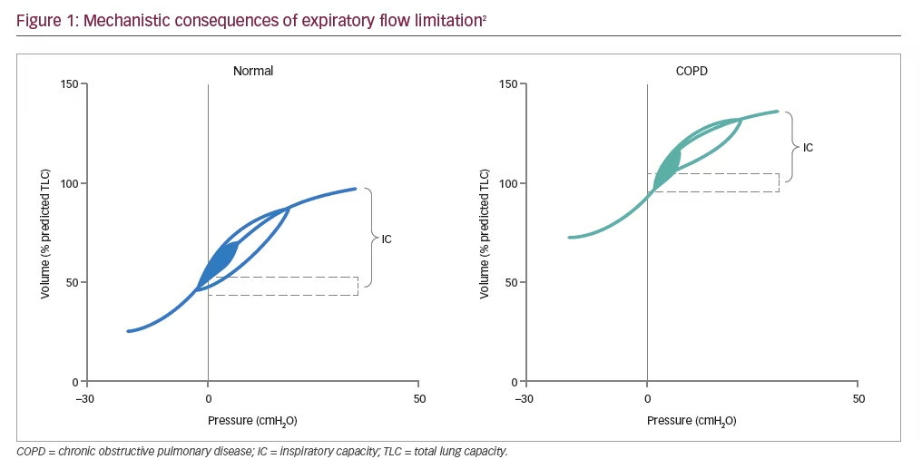
Tidal EFL in COPD arises because of the combined effects of reduced elastic lung recoil and increased airway resistance due to small airway inflammation, which causes a reduction in the luminal cross-sectional area.10,11 This, in turn, results in gas trapping on expiration10,12 and lung hyperinflation, a key mechanistic consequence of tidal EFL (Figure 1).2

In healthy subjects, EELV and inspiratory capacity (IC) are maintained throughout exercise.2 Any changes that occur in EELV as a result of obstructive lung disease are reflected by changes in IC, as total lung capacity (TLC [EELV+IC]) remains unchanged during exercise.2 In COPD, dynamic hyperinflation is the temporary and variable increase in EELV above its baseline value, which is aggravated further as expiratory time shortens, e.g., during exercise, resulting in further increases in EELV.2 Dynamic hyperinflation has been shown to be strongly associated with EFL during exhalation,10 which in mild COPD usually occurs during exercise.13 However, as the condition worsens EFL and dynamic hyperinflation can also be present at rest.13 EFL contributes independently to symptoms during COPD exacerbations (when compared to patients without EFL),14 the most distressing of which is dyspnoea (breathlessness).15 Patients with COPD have described dyspnoea as an unsatisfied inspiratory effort (i.e., they can’t get enough air in), having inspiratory difficulty or experiencing shallow breathing.2

New ways to non-invasively measure expiratory flow limitation in chronic obstructive pulmonary disease

Assessment of small airway function in clinical practice has remained a challenge for many years and progress in this area has been impeded by technical limitations related to the small size of the airways and to their location deep within the thoracic cavity.16 Several methods have been proposed to detect tidal EFL in patients with COPD, including the invasive oesophageal balloon technique, non-invasive abdominal compression, NEP and FOT.9,17,18 The challenges with some of these methods include the impact of patient position on reproducibility, e.g., seated or supine (abdominal compression),19 as well the subjective nature of the assessment (NEP), which has resulted in a reassessment of the utility of FOT.8

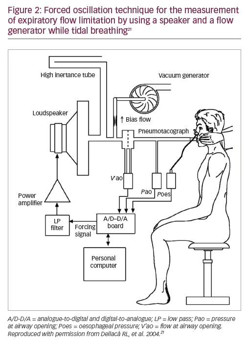
DuBois et al. first introduced FOT in 1956 (Figure 2),20 but it is only now gaining clinical recognition. It is an objective measure that does not require the performance of respiratory manoeuvres and offers a simple, detailed and repeatable approach to investigating the mechanical properties of the respiratory system.8,21,22 It has also been shown to be as sensitive as spirometry in detecting impairments of lung function.22 FOT is performed at higher frequencies than regular breathing (typically between 5–25 Hz).23,24 Reactance at 5 Hz during the expiratory phase (using the inspiratory reactance as a baseline) appears to be particularly sensitive to EFL, and this has been used in an effort to assess small airway function in patients with COPD.25,26

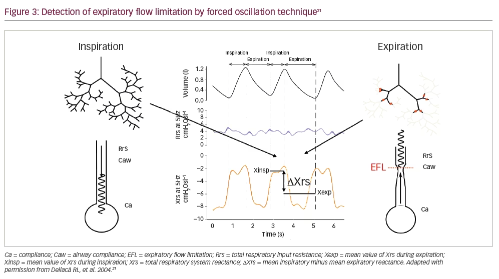
FOT can identify EFL when a large difference in within-breath inspiratory and expiratory reactance occurs due to the development of choke points throughout the bronchial tree (Figure 3).8,21 Measuring reactance over multiple breaths is preferable as it retrieves information from a longer time span than breath-by-breath measurements. As a result, aberrations in breathing pattern occurring within an individual breath are averaged over the entire measurement period, and so yield a more accurate measurement.7 Higher values are common in patients with COPD even versus smoking controls, with high values tending to remain consistent over time.7

Why is it important to assess expiratory flow limitation in chronic obstructive pulmonary disease?

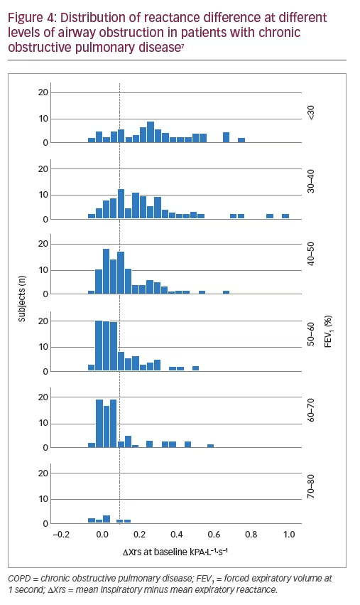
The presence of EFL during a COPD exacerbation has potential clinical implications, such as signalling the presence of more severe physiological perturbations that are not detectable by spirometry.14 However, EFL is often under-recognised in the clinical setting.27 EFL in patients with COPD is associated with lower forced expiratory volume in 1 second (FEV1) (Figure 4) and greater hyperinflation, as well as a significant correlation with worse health status, symptom score and burden of disease, as assessed by the St George’s Respiratory Questionnaire.10 Increased EFL index values at the time of hospital admission for a COPD exacerbation may signal more severe physiological perturbations not detectable by spirometry, since increased EFL has been shown to correlate with longer hospital stays.14

Detection of tidal EFL can also help to identify factors limiting exercise tolerance such as IC, as resting EFL clearly separates two populations of patients with significant differences in exercise tolerance.28,29 Tidal EFL has been shown to correlate with the degree of breathlessness7 and walking distance, and can be used to identify the subgroup of patients with COPD worse clinical outcomes.30 It is also important to assess EFL in clinical practice, as a decrease in index values may relate to improvements in symptoms during recovery from an exacerbation.14 Bronchodilators can improve dynamic airway function, allowing improved lung emptying with each breath, which is associated with improvements in dyspnoea as a result of increased IC and reduction in EELV.2 A significant reduction of dynamic hyperinflation in patients with EFL at rest after bronchodilator administration supports the importance of categorising patients with COPD into subgroups with and without EFL, in order to predict response to bronchodilator therapy.31,32

Identification or screening of EFL with FOT can also guide the use of continuous positive airway pressure or positive end expiratory pressure in mechanically ventilated patients with COPD, allowing targeting of the minimum pressure able to counteract the detrimental effects of intrinsic positive end expiratory pressure without unnecessarily affecting haemodynamics and/or impairing inspiratory muscle function by increasing operating volumes.33

Overall, EFL could be useful for stratifying patients by the severity of their COPD or treatment responses.4 The recognition and monitoring of EFL before and after exacerbation could potentially be used to guide or titrate treatment, reduce the duration of hospital stays and improve clinical outcomes for patients with COPD.30

Article Information:
Disclosure

Peter Calverley has nothing to disclose. Raffaele L Dellacà’s
institution, Politecnico di Milano University, licensed a patent on the
automatic detection of Expiratory Flow Limitation to Philips Respironics.
Symposium Faculty: Peter Calverley, Professor of Respiratory Medicine and
Honorary Consultant Respiratory Physician at Aintree Hospitals, Liverpool,
UK; Raffaele L Dellacà, Associate Professor, Politecnico di Milano University,
Milan, Italy; David White, Chief Scientific Officer, Philips Sleep & Respiratory
Care, Denver, CO, US; Robert Romano, Biomedical Engineer at Philips Medical
Systems, Philips Home Healthcare Solutions, Pittsburgh, PA, US.

Review Process

This article reports the proceedings of a sponsored
satellite symposium held at the ERCA-JIVD 3rd Joint International Meeting.
As such, it has not been subject to this journal’s usual peer-review process.
The report was reviewed for scientific accuracy by the symposium
speakers and a member of the editorial board before publication.

Authorship

All named authors meet the criteria of the International Committee of
Medical Journal Editors for authorship for this manuscript, take responsibility for the
integrity of the work and have given final approval for the version to be published.

Correspondence

Peter Calverley, Professor of Respiratory Medicine, Aintree University Hospital, Lower Lane, Liverpool, Merseyside, L9 7AL, UK. E: pmacal@liverpool.ac.uk

Support

Philips Respironics funded the publication of this article through an independent educational grant and were provided the opportunity to review the draft for medical accuracy. Any resulting changes were made at the expert reviewers’ discretion.

Access

This article is published under the Creative Commons Attribution Noncommercial
License, which permits any non-commercial use, distribution, adaptation
and reproduction provided the original author(s) and source are given appropriate credit.
© The Authors 2018.

Acknowledgements

Medical Writing support was provided by
Vanessa Lane, PhD, Touch Medical Communications, supported by
an independent educational grant from Philips Respironics.

Received

2018-07-16

References

  1. Vestbo J, Hurd SS, Agusti AG, et al. Global strategy for the diagnosis, management, and prevention of chronic obstructive pulmonary disease: GOLD executive summary. Am J Respir Crit Care Med. 2013;187:347–65.
  2. O’Donnell DE. Hyperinflation, dyspnea, and exercise intolerance in chronic obstructive pulmonary disease. Proc Am Thorac Soc. 2006;3:180–4.
  3. Hyatt RE. The interrelationships of pressure, flow, and volume during various respiratory manoeuvres in normal and emphysematous subjects. Am Rev Respir Dis. 1961;83:676–83.
  4. Mikamo M, Shirai T, Mori K, et al. Predictors of expiratory flow limitation measured by forced oscillation technique in COPD. BMC Pulmonary Med. 2014;14:23.
  5. Eltayara L, Becklake MR, Volta CA, et al. Relationship between chronic dyspnea and expiratory flow limitation in patients with chronic obstructive pulmonary disease. Am J Respir Crit Care Med. 1996;154:1726–34.
  6. Gagnon P, Guenette JA, Langer D, et al. Pathogenesis of hyperinflation in chronic obstructive pulmonary disease. Int J Chron Obstruct Pulmon. 2014:9:187–201.
  7. Aarli BB, Calverley PMA, Jensen RL, et al. Variability of within-breath reactance in COPD patients and its association with dyspnoea. Eur Respir J. 2015;45:625–34.
  8. Dellacà RL, Duffy N, Pompilio PP, et al. Expiratory flow limitation detected by forced oscillation and negative expiratory pressure. Eur Respir J. 2007;29:363–74.
  9. Baydur A. Expiratory flow limitation in intra and extrathoracic respiratory disorders: use of the negative expiratory pressure technique – review and recent developments. In: Oncogenesis inflammatory and parasitic tropical diseases of the lung. Chapter 6. Jean-Marie Kayembe (ed), InTech, 2013; DOI: 10.5772/53270.
  10. Dean J, Kolsum U, Hitchen P, et al. Clinical characteristics of COPD patients with tidal expiratory flow limitation. Int J Chron Obstruct Pulmon Dis. 2017;12:1503–6.
  11. Hogg JC, Chu F, Utokaparch S, et al. The nature of small-airway obstruction in chronic obstructive pulmonary disease. N Engl J Med. 2004;350:2645–53.
  12. Thomas M, Decramer M, O’Donnell DE. No room to breathe: the importance of lung hyperinflation in COPD. Prim Care Respir J. 2013;22:101–11.
  13. O’Donnell D, Revill S, Webb K. Dynamic hyperinflation and exercise intolerance in chronic obstructive pulmonary disease. Am J Respir Crit Care Med. 2001;164:770–7.
  14. Jetmalani K, Timmins S, Brown NJ, et al. Expiratory flow limitation relates to symptoms during COPD exacerbations requiring hospital admission. Int J Chron Obstruct Pulmon Dis. 2015;10:939–45.
  15. O’Donnell DE, Elbehairy AF, Faisal A, et al. Exertional dyspnoea in COPD: the clinical utility of cardiopulmonary exercise testing. Eur Respir Rev. 2016;25:333–47.
  16. Brusasco V, Barisione G, Crimi E. Pulmonary physiology: future directions for lung function testing in COPD. Respirology. 2015;20:209–18.
  17. Koulouris N, Valta P, Lavoie A, et al. A simple method to detect expiratory flow limitation during spontaneous breathing. Eur Respir J. 1995;8:306–13.
  18. Tantucci C. Expiratory flow limitation definition, mechanisms, methods, and significance. Pulm Med. 2013;2013:749860.
  19. Ninane V, Leduc D, Kafi SA, et al. Detection of expiratory flow limitation by manual compression of the abdominal wall. Am J Respir Crit Care Med. 2001;163:1326–30.
  20. DuBois AB, Brody AW, Lewis DH, Burgess BF Jr. Oscillation mechanics of lungs and chest in man. J Appl Physiol. 1956;8:587–94.
  21. Dellacà RL, Santus P, Aliverti A, et al. Detection of expiratory flow limitation in COPD using the forced oscillation technique. Eur Respir J. 2004;23:232–40.
  22. Oostveen E, MacLeod D, Lorino H, et al. The forced oscillation technique in clinical practice: methodology, recommendations and future developments. Eur Respir J. 2003;22:1026–41.
  23. Verbanck S. Physiological measurement of the small airways. Respiration. 2012;84:177–88.
  24. Dellacà R, Kaczka DW. Oscillation mechanics of the respiratory system: applications to lung disease. Crit Rev Biomed Eng. 2011;39:337–59.
  25. Kanda S, Fujimoto K, Komatsu Y, et al. Evaluation of respiratory impedance in asthma and COPD by an impulse oscillation system. Intern Med. 2010;49:23–30.
  26. Paredi P, Goldman M, Alamen A, et al. Comparison of inspiratory and expiratory resistance and reactance in patients with asthma and chronic obstructive pulmonary disease. Thorax. 2010;65:263–7.
  27. Junhasavasdikul D, Telias I, Grieco DL, et al. Expiratory flow limitation during mechanical ventilation. Chest. 2018; DOI: 10.1016/j.chest.2018.01.046 [Epub ahead of print].
  28. Diaz O, Villafranca C, Ghezzo H, et al. Role of inspiratory capacity on exercise tolerance in COPD patients with and without tidal expiratory flow limitation at rest. Eur Respir J. 2000;16:269–75.
  29. Koulouris NG, Kaltsakas G, Palamidas AF, Gennimata SA. Methods for assessing expiratory flow limitation during tidal breathing in COPD patients. Pulm Med. 2012;2012:234145.
  30. Aarli BB, Calverley PMA, Jensen RL, et al. The association of tidal EFL with exercise performance, exacerbations, and death in COPD. Int J Chron Obstruct Pulmon. 2017;12:2179–88.
  31. Tantucci C, Duguet A, Similowski T, et al. Effect of salbutamol on dynamic hyperinflation in chronic obstructive pulmonary disease patients. Eur Respir J. 1998;12:799–804.
  32. Dellacà RL, Pompilio PP, Walker PP, et al. Effect of bronchodilation on expiratory flow limitation and resting lung mechanics in COPD. Eur Respir J. 2009;33:1329–37.
  33. Dellacà RL, Rotger M, Aliverti A, et al. Noninvasive detection of expiratory flow limitation in COPD patients during nasal CPAP. Eur Respir J. 2006;27:983–91.

Further Resources

Share this Article
Related Content In COPD
  • Copied to clipboard!
    accredited arrow-down-editablearrow-downarrow_leftarrow-right-bluearrow-right-dark-bluearrow-right-greenarrow-right-greyarrow-right-orangearrow-right-whitearrow-right-bluearrow-up-orangeavatarcalendarchevron-down consultant-pathologist-nurseconsultant-pathologistcrosscrossdownloademailexclaimationfeedbackfiltergraph-arrowinterviewslinkmdt_iconmenumore_dots nurse-consultantpadlock patient-advocate-pathologistpatient-consultantpatientperson pharmacist-nurseplay_buttonplay-colour-tmcplay-colourAsset 1podcastprinter scenerysearch share single-doctor social_facebooksocial_googleplussocial_instagramsocial_linkedin_altsocial_linkedin_altsocial_pinterestlogo-twitter-glyph-32social_youtubeshape-star (1)tick-bluetick-orangetick-red tick-whiteticktimetranscriptup-arrowwebinar Sponsored Department Location NEW TMM Corporate Services Icons-07NEW TMM Corporate Services Icons-08NEW TMM Corporate Services Icons-09NEW TMM Corporate Services Icons-10NEW TMM Corporate Services Icons-11NEW TMM Corporate Services Icons-12Salary £ TMM-Corp-Site-Icons-01TMM-Corp-Site-Icons-02TMM-Corp-Site-Icons-03TMM-Corp-Site-Icons-04TMM-Corp-Site-Icons-05TMM-Corp-Site-Icons-06TMM-Corp-Site-Icons-07TMM-Corp-Site-Icons-08TMM-Corp-Site-Icons-09TMM-Corp-Site-Icons-10TMM-Corp-Site-Icons-11TMM-Corp-Site-Icons-12TMM-Corp-Site-Icons-13TMM-Corp-Site-Icons-14TMM-Corp-Site-Icons-15TMM-Corp-Site-Icons-16TMM-Corp-Site-Icons-17TMM-Corp-Site-Icons-18TMM-Corp-Site-Icons-19TMM-Corp-Site-Icons-20TMM-Corp-Site-Icons-21TMM-Corp-Site-Icons-22TMM-Corp-Site-Icons-23TMM-Corp-Site-Icons-24TMM-Corp-Site-Icons-25TMM-Corp-Site-Icons-26TMM-Corp-Site-Icons-27TMM-Corp-Site-Icons-28TMM-Corp-Site-Icons-29TMM-Corp-Site-Icons-30TMM-Corp-Site-Icons-31TMM-Corp-Site-Icons-32TMM-Corp-Site-Icons-33TMM-Corp-Site-Icons-34TMM-Corp-Site-Icons-35TMM-Corp-Site-Icons-36TMM-Corp-Site-Icons-37TMM-Corp-Site-Icons-38TMM-Corp-Site-Icons-39TMM-Corp-Site-Icons-40TMM-Corp-Site-Icons-41TMM-Corp-Site-Icons-42TMM-Corp-Site-Icons-43TMM-Corp-Site-Icons-44TMM-Corp-Site-Icons-45TMM-Corp-Site-Icons-46TMM-Corp-Site-Icons-47TMM-Corp-Site-Icons-48TMM-Corp-Site-Icons-49TMM-Corp-Site-Icons-50TMM-Corp-Site-Icons-51TMM-Corp-Site-Icons-52TMM-Corp-Site-Icons-53TMM-Corp-Site-Icons-54TMM-Corp-Site-Icons-55TMM-Corp-Site-Icons-56TMM-Corp-Site-Icons-57TMM-Corp-Site-Icons-58TMM-Corp-Site-Icons-59TMM-Corp-Site-Icons-60TMM-Corp-Site-Icons-61TMM-Corp-Site-Icons-62TMM-Corp-Site-Icons-63TMM-Corp-Site-Icons-64TMM-Corp-Site-Icons-65TMM-Corp-Site-Icons-66TMM-Corp-Site-Icons-67TMM-Corp-Site-Icons-68TMM-Corp-Site-Icons-69TMM-Corp-Site-Icons-70TMM-Corp-Site-Icons-71TMM-Corp-Site-Icons-72